- Get link
- X
- Other Apps
If these pockets are larger than 2. Research shows that there is an association between choroid plexus cysts and aneuploidy.
Https Mydoctor Kaiserpermanente Org Ncal Images Gen Us 20cpc 20handout Tcm63 15061 Pdf
What are choroid plexus cysts.

Choroid plexus cyst. The choroid plexus is the part of the brain that makes cerebrospinal fluid the fluid that normally bathes and protects the brain and spinal column. We present a case of a female patient with an incidental finding of bilateral choroid plexus cysts. 2152021 A choroid cyst is a small buildup of fluid in the choroid plexus a structure in the brain which produces cerebrospinal fluid CSF.
No other abnormalities were detected. These glands make the. Lopez JA1 Reich D.
More than 90 resolve by 26 weeks. In 1 to 2 of babies a cyst- a small round fluid filled area- is formed in the choroid plexus. Choroid Plexus Cyst A choroid plexus cyst is a small area of fluid that collects in a part of the brain called the choroid plexus.
An isolated choroid plexus cyst was not associated with a trisomy or other abnormalities in this study. The choroid plexuses are structures of the brain that make the cerebrospinal fluid the fluid that normally flows around the brain and spine. The choroid plexus is not part of the brain involved in thinking or development.
The choroid plexus is a spongy pair of glands located on each side of the brain. On evaluation the fetus was incidentally detected to have a right-sided choroid plexus cyst CPC of 5 mm. Choroid plexus cysts CPCs are incidental findings on sonograms of the neonatal head.
Ashish Jain Bijaylaxmi Behera. To identify a chroid plexus cyst CPC it must be imaged in two orthogonal planes and be greater than or equal to 3 mm in size. When a choroid plexus cyst was associated with an additional risk factor 105 of the patients had an abnormality.
A 29-year-old gravida 2 para 1 underwent antenatal ultrasonography USG at 18 weeks of gestation. This fluid bathes and protects the brain and spinal column. In about 1 to 2 percent of normal babies 1 out of 50 to 100 a tiny bubble of fluid is pinched off as the choroid plexus forms.
Platt LD Carlson DE Medearis AL Walla CA 1991 Fetal choroid plexus cysts in the second trimester of pregnancya cause for concern. Sometimes fluid becomes trapped and forms pockets in the choroid plexus. A normal finding on ultrasound Within a period of 25 years cystic structures in the choroid plexus were encountered at cerebral sonography in 70 neonates and babies 45 male 25 female.
Their prevalence in patients examined during the first 4 weeks of life n 55 was 3. How does it happen. Located on the left and the right side of the brain the choroid plexus is a gland that produces cerebrospinal fluid.
What is a Choroid plexus cyst. The incidence of CPCs and their association with childhood neurodevelopmental outcome remains unclear. Pilling D Sprigg A Abernethy L Walkinshaw S 1992 Isolated choroid plexus cysts-no grounds for complacency.
1Wyckoff Heights Medical Center Brooklyn NY 11237 USA. Amniocentesis is recommended when a choroid plexus cyst is found in association with additional risk factors. What is a Choroid Plexus Cyst.
Choroid cysts are most commonly identified as an ultrasound finding and are in fact not uncommon being. In most instances these are a normal variant. 3252019 Choroid Plexus Cysts CPC are small fluid filled areas in the brain and they are a common ultrasound finding in the fetus during the 2nd trimester of pregnancy.
Family physicians treating prenatal patients should understand the management of this sonographic finding. Single or multiple cystic areas 2 mm in diameter in one or both choroid plexuses of the lateral cerebral ventricles. 482020 Choroid plexus cysts CPC can be symptomatic if they are large in size andor cause obstructive hydrocephalus 13.
Associated with increased risk for trisomy 18 and possibly trisomy 21. Histologically most of these cysts prove to be degenerative xanthogranulomas XG.
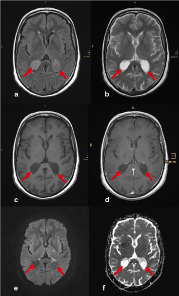
 Bilateral Choroid Plexus Cysts Xanthogranulomas
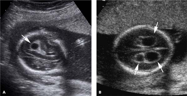
 Prenatal Ultrasound Findings A Choroid Plexus Cysts At 18 Weeks Of Download Scientific Diagram
 Wk 6 L 2 Choroid Plexus Cyst Radiology Case Radiopaedia Org Diagnostic Medical Sonography Plexus Products Radiology
 Sagittal Plane Of The Fetal Head Shows A Choroid Plexus Cyst Download Scientific Diagram
 Chromosomal Anomalies Radiology Key
 Isolated Fetal Choroid Plexus Cysts
 Isolated Large Bilateral Choroid Plexus Cysts Associated With Trisomy 18 Bmj Case Reports
 Choroid Plexus Anomalies Cysts And Papillomas Sciencedirect
 Choroid Plexus Cyst Radiology Case Radiopaedia Org
 Choroid Plexus Cyst Gynaecologia
 Choroid Plexus Cyst Cpc Hkog Info


Comments
Post a Comment